




Nasal Skin Necrosis or Blackening of Nasal Skin
Dr. Hamidreza Hosnani
ENT Specialist | Rhinoplasty Surgeon in Tehran | Nose Surgery Specialist
Necrosis means the death of living nasal tissue. The extent of necrosis damage depends on the size and amount of necrotic tissue. Small pinpoint areas of necrosis usually do not cause harm, but if a large amount of tissue becomes necrotic and the nasal framework is significantly damaged, it can affect the external appearance of the nose.
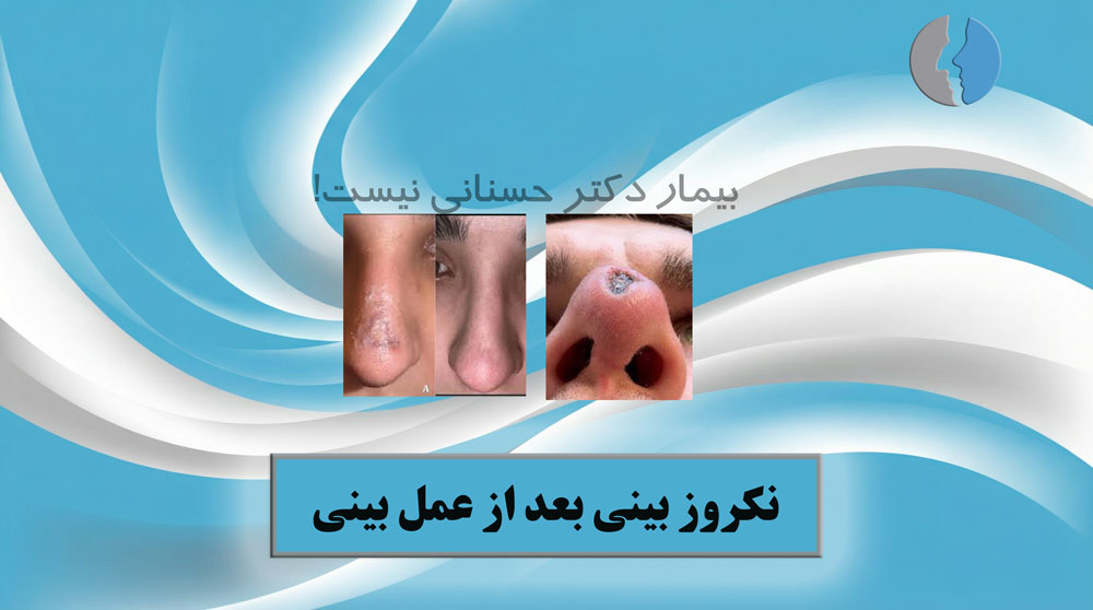
Nasal necrosis after surgery is a very rare complication that occurs when blood supply to a portion of the nasal skin or tissue is disrupted. This problem may be associated with symptoms such as skin discoloration, blisters, unusual pain, or wounds, and it is more likely to occur in situations such as excessive pressure on the skin, smoking, vascular disorders, or very aggressive surgery. However, according to Dr. Hosnani’s experience, when proper surgical principles are followed, blood supply is preserved, and postoperative care is appropriate, the likelihood of this complication is extremely low.
Navigating Post-Rhinoplasty Complications
Understanding the Risks
Nasal necrosis represents one of the most critical, albeit exceedingly rare, complications following rhinoplasty. While modern surgical techniques have significantly minimized these occurrences, patient awareness remains vital. Alongside necrosis, patients frequently inquire about localized depressions or irregularities in the nasal profile. It is essential to distinguish between tissue necrosis—a biological breakdown—and simple structural irregularities.Clinical Significance
Understanding the distinction between these conditions allows patients to maintain a realistic outlook on their recovery trajectory. While necrosis requires immediate medical intervention to prevent significant deformity, minor irregularities are often part of the healing process.Understanding Nasal Necrosis
Physiological Basis
Necrosis is characterized by the death of living cells, typically resulting from compromised blood supply. When oxygenated blood fails to reach the skin or underlying tissue, localized ischemia leads to cell death. This can be induced by external pressure, severe infection, or vascular damage during surgery.Severity and Classification
The clinical impact depends on the extent of the damage. Superficial necrosis might manifest as minor crusting that resolves with topical care. In contrast, extensive tissue loss involving the cartilaginous framework can necessitate complex reconstructive efforts to address visible depressions or structural perforation.Identifying Early Warning Signs
Cutaneous Changes
Patients must vigilantly monitor the skin's surface. Notable warning signs include persistent discoloration—such as dark gray, purple, or black patches—that does not resolve with standard post-operative cooling methods.Sensory Indicators
Beyond visual changes, patients should be alert to severe, localized burning sensations or uncharacteristic pain that worsens rather than improves over time. Foul-smelling discharge from suture lines is another critical indicator.According to Dr. Hosnani, early detection is the cornerstone of successful management. Patients should never hesitate to report sudden, intense pain or skin color changes during the first few weeks, as rapid intervention is the most effective safeguard against irreversible damage.
Etiology of Post-Surgical Complications
External and Systemic Influences
Several factors contribute to the risk profile of necrosis. Improperly applied splints can exert excessive force, leading to local hypoxia. Furthermore, systemic conditions such as uncontrolled diabetes or vascular diseases can significantly impair the body's natural wound-healing mechanisms.Lifestyle Factors
Lifestyle choices are paramount. The use of nicotine—including cigarettes and vaping—is a major vasoconstrictor that restricts blood flow to the distal tissues of the nose, significantly increasing the likelihood of vascular compromise.Addressing Post-Operative Depressions
Temporary Edema versus Structural Issues
Not all irregularities signify necrosis. In the early stages, depressions are often the result of uneven swelling, skin retraction, or the specific way the splint sits on the nasal bridge. These are frequently amenable to conservative treatments.The Six-Month Assessment
When irregularities persist beyond six months, it may indicate a structural deficiency. As experienced surgeons like Dr. Hosnani advise:It is common for patients to worry about nasal contours during the first few months. However, the nose undergoes constant, subtle changes throughout the first year. We generally refrain from definitive structural assessment until the tissues have fully matured, typically after twelve months.
Therapeutic Interventions
Non-Surgical Bridge Treatments
For patients experiencing significant aesthetic distress during the waiting period, temporary dermal fillers may be utilized. These standard, biocompatible agents are injected to improve symmetry, offering a bridge to the one-year mark without permanent modification of the skeletal structure.Surgical Reconstruction
Once the recovery timeline reaches one year, if structural depressions remain, secondary rhinoplasty may be considered. This procedure often involves utilizing graft tissues, such as septal, ear, or rib cartilage, to reinforce the nasal architecture.Mitigating Risks
Proactive Patient Care
Reducing the risk of complications requires a collaborative approach between the patient and the surgical team. This includes adherence to post-operative protocols, such as strict avoidance of nicotine and careful management of nasal dressings.Long-Term Monitoring
Regular follow-up appointments allow the surgeon to monitor the healing progression effectively. According to the experience of Dr. Hosnani:The path to aesthetic stability is paved with patience and precise aftercare. By choosing an experienced surgeon and strictly following recovery guidelines, patients can minimize risks and ensure that their final result is both structurally sound and aesthetically pleasing.