




Feeling of Adhesions Inside the Nose
Dr. Hamidreza Hosnani
ENT Specialist | Rhinoplasty Surgeon in Tehran | Nose Surgery Specialist
Internal nasal adhesions are one of the functional complications of rhinoplasty and are caused by the failure of surgical incisions and wounds to heal properly and the sticking of internal nasal tissues. To diagnose the cause of nasal adhesions, sometimes a CT scan is required. The presence of internal nasal adhesions and internal nasal deviation can cause congestion and breathing problems. Rarely, the sense of smell may be disturbed due to internal nasal adhesions, which of course is temporary.
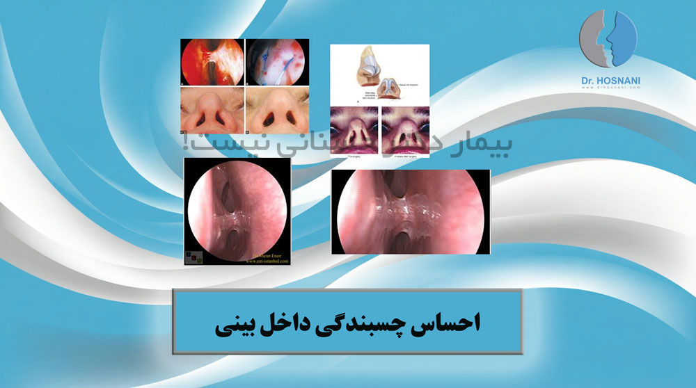
Internal nasal adhesion, or “synechia,” usually occurs when internal nasal surfaces stick together during the healing process. This condition can cause persistent nasal obstruction, difficulty breathing, a whistling sound during breathing, or even complete blockage of one side of the nose, and it typically appears several weeks to several months after surgery.
Intranasal Adhesions: Understanding Functional Obstruction after Surgery
Intranasal adhesion, clinically referred to as synechia, is a recognized functional complication that may arise following aesthetic or reconstructive rhinoplasty. This condition occurs when opposing mucosal surfaces within the nasal cavity undergo an irregular healing process, leading to the formation of fibrous bridges between structures that should remain separate, such as the nasal sidewall and the septum.Pathophysiology and the Healing Process
The primary cause of these adhesions is the disruption of the delicate mucosal lining. During the recovery phase, if two raw or inflamed mucosal surfaces remain in contact, they may fuse together. This unintended biological union creates a physical barrier that restricts the natural flow of air through the nasal passages.Diagnostic Methods and Specialized Imaging
Identifying an adhesion requires a systematic clinical approach. When a patient reports chronic congestion or a persistent sensation of blockage in one or both nostrils, a thorough evaluation is necessary. While many adhesions are visible during a standard physical exam, some are located deeper within the nasal vault. In such cases, specialized imaging, such as a CT scan of the sinuses and nasal passages, is utilized to provide a detailed map of the internal anatomy.
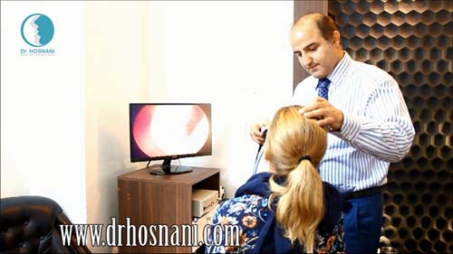
Dr. Hosnani examining Esmeralda’s nose before surgery. Preoperative nasal examination is a vital step during which the surgeon evaluates the internal and external structure of the nose, the degree of septal deviation, skin thickness, cartilage quality, and the harmony of the nose with other facial features. This evaluation helps identify possible breathing problems and allows the surgeon to design a surgical plan tailored to both aesthetic and functional needs.
Clinical Consequences of Nasal Tissue Bonding
Impact on Respiratory Dynamics and Sleep
Intranasal adhesions can significantly impair the quality of life by causing chronic airway obstruction. When the air passage is narrowed by scar tissue, the patient may experience difficulty breathing through the nose, especially during physical exertion or sleep. This can lead to secondary issues such as mouth breathing, dry throat, and a measurable decrease in overall sleep quality.Sensory Disturbances and Olfactory Health
Although less common, some patients may notice a temporary reduction in their sense of smell. This is typically not a neurological deficit but rather a physical blockage preventing scent molecules from reaching the olfactory region at the roof of the nasal cavity. Resolving the adhesion often restores the natural airflow dynamics and sensory function. According to the clinical observations of Dr. Hosnani:"Intranasal adhesions act as a physical dam within the respiratory stream. Restoring the sense of smell in these patients is often as simple as surgically releasing these mucosal bridges to allow air to reach the olfactory epithelium once again."
Advanced Treatment Modalities for Adhesion Resolution
The Use of Physical Barriers and Splints
Treating intranasal adhesions involves more than just a simple release of the tissue. To prevent the surfaces from bonding again during the new healing phase, surgeons often employ internal nasal splints or spacers. These medical-grade barriers act as a physical shield, ensuring that the healing mucosal walls remain separated until they have completely epithelialized.Structural Correction via Cartilage Grafting
The location of the adhesion dictates the surgical strategy. If the bonding occurs at the internal nasal valve—the narrowest part of the airway—the treatment often involves more than just tissue release. To prevent a functional collapse of the valve, surgeons may use cartilage grafts to reinforce the area. Based on the experience of Dr. Hosnani:"When adhesions compromise the internal valve, structural integrity is paramount. We often utilize spreader grafts or other cartilaginous supports to ensure that once the adhesion is cleared, the airway remains wide and functionally stable."
Foundation Correction and the Importance of Examination
Addressing Residual Septal Deviation
In cases where adhesions form lower in the nasal cavity, the underlying cause is often an uncorrected septal deviation. If the septum remains tilted toward the lateral wall, it creates a narrow gap that is highly prone to mucosal contact and subsequent scarring. In these scenarios, the fundamental treatment is the surgical correction of the septum to increase the distance between internal structures.
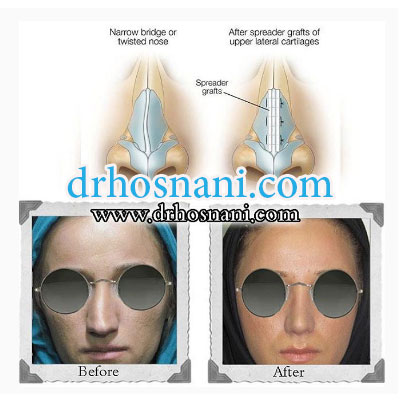
Noses that have a narrow bridge or are crooked and twisted are corrected using spreader grafts placed along the upper lateral cartilages. In this patient, the crooked and twisted nasal septum was repaired using a spreader graft. Grafts are materials used to strengthen the nasal framework or to correct nasal deviation. Dr. Hosnani usually uses grafts taken from the patient’s own body, known as autografts. In this patient, septal cartilage was used as the graft.
The Necessity of a Physical Consultation
Ultimately, a definitive diagnosis and a personalized treatment plan can only be established through a direct clinical examination. Because every patient's internal anatomy and healing response are unique, a specialist must visualize the interior of the nose to determine the exact origin and severity of the adhesion. As experienced surgeons like Dr. Hosnani emphasize:"Digital images and patient descriptions are helpful, but an endoscopic evaluation is the gold standard. It allows us to differentiate between simple mucosal bridges and more complex structural obstructions, ensuring the chosen intervention is effective and permanent."

Diagnosis of a Deviated Septum or Nasal Polyps Is Only Possible Through Examination