


What Is a Nasal CT Scan?
Dr. Hamidreza Hosnani
ENT Specialist | Rhinoplasty Surgeon in Tehran | Nose Surgery Specialist
A CT scan image of the internal structure of the nose is usually taken by the patient before rhinoplasty and presented to the surgeon. During surgery, these images are placed on an illuminated viewing panel mounted on the wall in the operating room so the doctor can consider the internal nasal anatomy while performing the procedure.

For further evaluation of internal nasal lesions that require more investigation, diagnostic tools such as CT scans can be helpful.
One of the best methods for this evaluation is a CT scan. A CT scan can provide very detailed, multi‑dimensional images of the bones, the nasal septum, and the sinuses, helping the surgeon assess the complete condition of the nose before surgery.
However, a CT scan is not necessary for everyone who plans to undergo rhinoplasty and is usually recommended only in certain situations.
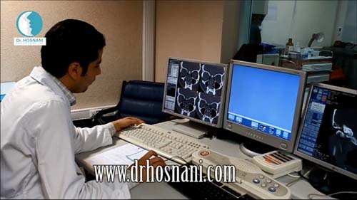
Laboratory Technician Preparing CT Scan Images
A CT scan is an advanced imaging method that uses X‑rays to capture multiple cross‑sectional images of the nose and sinuses. These images are then combined so the physician can compare them with the findings from the physical examination.
Using this advanced imaging technique, many conditions inside the nose and sinuses can be diagnosed more accurately.
Problems such as a deviated septum, nasal polyps, and infections of the sinuses (sinusitis) can be precisely diagnosed with a CT scan of the nose and sinuses.

A CT scan before rhinoplasty is usually requested when the surgeon needs a more detailed evaluation of the internal structures of the nose and sinuses, especially in patients with a history of nasal obstruction, severe septal deviation, polyps, or breathing problems. This imaging method provides detailed cross-sectional and three-dimensional views of the bones, nasal septum, and sinuses, helping the surgeon plan more accurately for correcting deviations and internal nasal problems at the same time as cosmetic surgery. However, it is not necessary for all patients and is often recommended only in specific cases.
What Is a Nasal CT Scan?
A CT scan is an advanced imaging method that uses X‑rays and computer processing to create highly detailed cross‑sectional images of the internal structures of the body.In a CT scan of the nose and sinuses, clear images can be obtained of:
◆ Nasal bones
◆ The nasal septum
◆ Nasal turbinates
◆ The surrounding sinuses
◆ Sinus drainage pathways
◆ The presence of polyps or inflammation
These images allow the physician to examine the internal structure of the nose in a highly detailed three‑dimensional view.

After the blood test, the patient must go to medical imaging centers to obtain CT scans. CT scan images are necessary to be displayed in the operating room and increase the surgeon's accuracy during surgery.
Why Might a CT Scan Be Requested Before Rhinoplasty?
In some patients, a simple examination or nasal endoscopy may not be enough to diagnose the problem accurately. In such cases, a CT scan can provide more comprehensive information for the surgeon.The main reasons for requesting a CT scan before rhinoplasty include:
◆ Detailed evaluation of a deviated septum
◆ Assessment of sinus condition
◆ Diagnosis of nasal polyps
◆ Investigating the cause of chronic nasal obstruction
◆ Identifying complex structural problems
◆ Planning combined sinus and nasal surgery

On the Day of Surgery, Your CT Scan Images Are Installed on the Wall Above You and the Doctor Reviews Them Before Surgery
Which Patients Are More Likely to Need a CT Scan Before Rhinoplasty?
1. People With Chronic Nasal Obstruction
If a person has long‑term blockage in one or both sides of the nose, the doctor may request a CT scan to determine the cause.This obstruction may be caused by:
◆ Severe deviation of the nasal septum
◆ Enlarged nasal turbinates
◆ Nasal polyps
◆ Sinus inflammation
A CT scan can identify the exact location of these problems.

CT Scan Device Analyzing the Nose
2. Patients With Severe Septal Deviation
A deviated nasal septum is one of the most common conditions that can cause:◆ Nasal obstruction
◆ Snoring during sleep
◆ Difficulty breathing
In severe cases, a CT scan helps the surgeon determine the exact location and degree of the deviation and design a more precise surgical plan.

Receiving the CT Scan Test Result
3. People With a History of Chronic Sinusitis
Patients who suffer from recurrent or chronic sinusitis often require a detailed examination of the sinuses before rhinoplasty.In these cases, a CT scan can show:
◆ Which sinuses are affected
◆ Whether the sinus drainage pathways are blocked
◆ Whether sinus surgery is needed at the same time as rhinoplasty
In some cases, the doctor may perform endoscopic sinus surgery (FESS) simultaneously with rhinoplasty.

Nasal and Sinus Lesions Can Be Diagnosed with CT Scan
4. Presence of Nasal Polyps
Nasal polyps are soft, non‑cancerous growths that develop due to chronic inflammation of the nasal lining. They can cause:◆ Severe nasal obstruction
◆ Reduced sense of smell
◆ Post‑nasal drip

Dr. Hosnani Performing Rhinoplasty. The Patient’s CT Scan Images Are Installed on the Wall Above the Head
◆ Determine the exact location of the polyps
◆ Evaluate their extent
◆ Choose the most appropriate treatment method