



What Is a Nasal Turbinate?
Dr. Hamidreza Hosnani
ENT Specialist | Rhinoplasty Surgeon in Tehran | Nose Surgery Specialist
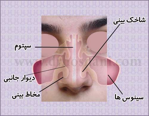
The nasal turbinates are bony structures covered with soft tissue (mucosa) located along the inner walls of the nose. There are three pairs: inferior, middle, and superior. Their main function is to humidify, warm, and filter the air we breathe. If these turbinates enlarge due to allergies or inflammation, they can cause nasal obstruction and breathing difficulty. In such cases, turbinate reduction surgery (turbinoplasty) may be performed.
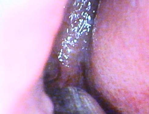
Enlarged Nasal Turbinates May Remain as a Complication When Surgery Is Performed by Inexperienced Surgeons
Understanding Nasal Turbinates and Their Role in Respiration
The **nasal turbinates**, medically referred to as turbinates or conchae, are bone structures coated with a specialized mucosal lining located within the nasal cavity. These structures perform vital functions in regulating airflow, humidifying the air, warming it to body temperature, and filtering inhaled particulates before they reach the lungs.
Contrary to misconceptions that they are vestigial or unnecessary features, turbinates are among the most essential components of the upper respiratory system. Their health and proper function have a direct impact on the overall quality of one’s breathing.
The Anatomy and Location of Nasal Turbinates
Anatomical Positioning
Turbinates are situated along the lateral walls inside the nose, appearing as curved, shelf-like projections. By increasing the surface area through which inhaled air must pass, these structures optimize the efficiency of the respiratory process.
Categorization of Turbinates
In each nasal passage, there are typically three turbinates:
• The Inferior Turbinate: The largest and most significant structure regarding the regulation of total airflow.
• The Middle Turbinate: Plays a crucial role in the drainage pathways of the sinuses.
• The Superior Turbinate: A smaller structure located in closer proximity to the olfactory region.
• The Middle Turbinate: Plays a crucial role in the drainage pathways of the sinuses.
• The Superior Turbinate: A smaller structure located in closer proximity to the olfactory region.
Clinical Impact of the Inferior Turbinate
The inferior turbinate exerts the greatest influence on the sensation of nasal patency, as its swelling is the most common cause of significant airway obstruction.
In Dr. Hosnani’s view, the turbinates are not merely anatomical obstacles but are dynamic physiological regulators that must be treated with absolute precision during any surgical intervention.
Physiological Functions of Turbinates
Air Filtration and Conditioning
The mucosa enveloping the turbinates contains a rich network of blood vessels. These vessels facilitate the warming of cold inhaled air, ensuring it is conditioned before reaching the lower airways. Simultaneously, the mucosal secretions serve to trap dust, allergens, and microorganisms, effectively cleansing the inhaled air.
Defense Mechanisms
Beyond conditioning, this mucosal layer provides a biological barrier. Consequently, the indiscriminate removal or severe damage to these tissues can significantly disrupt the nose's natural ability to protect the respiratory tract.
As observed by Dr. Hosnani, preserving the functional integrity of the mucosal layer is paramount; therefore, expert practitioners like Dr. Hosnani prioritize conservative surgical approaches to maintain these physiological defenses.
Nasal Turbinate Hypertrophy
Defining the Condition
Turbinate **hypertrophy** refers to the chronic enlargement or swelling of the nasal turbinates, which can result in a partial or complete blockage of the airflow pathways.
Etiology of Enlargement
The most significant factors contributing to this condition include:
• Seasonal or perennial allergies
• Chronic rhinitis (persistent inflammation of the nasal mucosa)
• Environmental irritants, such as tobacco smoke
• Deviated nasal septum (causing compensatory enlargement of the opposite turbinate)
• Chronic rhinitis (persistent inflammation of the nasal mucosa)
• Environmental irritants, such as tobacco smoke
• Deviated nasal septum (causing compensatory enlargement of the opposite turbinate)
Rebound Congestion
In certain individuals, the prolonged use of over-the-counter decongestant sprays can lead to a rebound effect, causing the turbinates to swell even further.
Symptomatic Presentation
Patients experiencing **nasal obstruction** due to turbinate hypertrophy frequently report:
• A sensation of persistent nasal blockage
• Difficulty breathing through one or both nostrils
• Nocturnal snoring
• A reduced sense of smell in advanced cases
• Difficulty breathing through one or both nostrils
• Nocturnal snoring
• A reduced sense of smell in advanced cases
Diagnostic Approaches
Clinical Examination and Endoscopy
An accurate diagnosis of turbinate enlargement is conducted by an otolaryngologist. Using a nasal speculum or an endoscope, the physician can directly visualize the interior of the nasal cavity. Nasal endoscopy, in particular, allows for a more comprehensive inspection of the turbinates, the septum, and the sinus ostia.
Differentiation from Septal Deviation
While a septal deviation involves a curvature of the central cartilage, turbinate hypertrophy involves the swelling of the lateral nasal walls. It is not uncommon for both issues to coexist, requiring a dual approach to fully restore optimal airway patency.
Treatment Modalities
Pharmacological Interventions
In mild to moderate cases, treatment may involve intranasal corticosteroid sprays, antihistamines, or regular saline irrigation to reduce mucosal inflammation. Such treatments generally represent the first line of defense, particularly for patients with underlying allergic conditions.
Surgical Volume Reduction
If conservative treatments fail to provide relief, surgical volume reduction is indicated. Modern techniques, such as conchoplasty, focus on reducing excess tissue while preserving as much of the natural mucosal function as possible. The primary goal of this surgery is to optimize the airway without compromising the nose's physiological performance.
Based on Dr. Hosnani’s clinical experience, the decision to proceed with surgery is made only after a thorough evaluation of the patient’s specific anatomy and symptom profile.
Considerations in Rhinoplasty
Combined Functional and Cosmetic Approaches
Many patients present with concurrent functional and aesthetic concerns. If turbinate hypertrophy is present, the surgeon can perform volume reduction simultaneously with rhinoplasty, ensuring both appearance and breathing are addressed in one operation.
Impact on Post-Operative Quality of Life
When performed correctly, this combined approach can significantly enhance respiratory function, preventing issues that might otherwise arise after cosmetic surgery.
Risks of Excessive Resection
Resecting excessive turbinate tissue carries a risk of a rare and complex complication known as **empty nose syndrome**. In this state, despite the airway being physically open, the patient experiences paradoxical sensations of breathlessness or severe nasal dryness. Consequently, conservative strategies that prioritize mucosal preservation are preferred by seasoned specialists such as Dr. Hosnani.
Reflecting Dr. Hosnani’s clinical expertise, the enduring success of nasal surgery is defined not only by the cosmetic outcome but by the preservation of functional breathing throughout the recovery period.
Post-Operative Recovery and Insights
Recovery Expectations
Recovery following turbinate surgery is typically faster than that of a comprehensive rhinoplasty. Consistent use of saline irrigation, avoidance of irritants like tobacco smoke, and adherence to professional guidance are essential for minimizing mucosal swelling and accelerating the healing process.
Timeline for Improvement
For the majority of patients, significant improvement in breathing is realized within a few weeks as the initial post-operative swelling subsides. The final outcome is achieved once the mucosal tissue has fully remodeled.
Recurrence and Long-Term Outcomes
If underlying factors like severe allergies persist, there remains a possibility of partial swelling recurrence. Managing the underlying condition is crucial for long-term success.
Turbinates versus Nasal Polyps
It is vital to distinguish that turbinates are natural structures, whereas **nasal polyps** are abnormal, inflammatory masses resulting from chronic mucosal disease. The nature of these conditions and their respective treatments are entirely distinct.